

Pig Anatomy and Terminology
"Pigtionary"
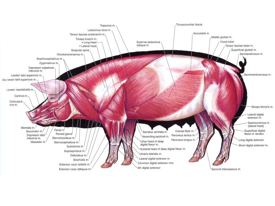
Muscular System
There are three types of muscle in the pig:
Terminology
Asymmetric hind quarter syndrome - One hind leg muscle mass appears less than the other. It can arise where poor quality iron injections are given or it may be a congenital condition. It may be part of the porcine stress syndrome (PSS).
Back muscle necrosis - Sudden acute lameness and swellings of the lumber muscle often associated with PSS.
Congenital muscle hypertrophy - A breeding defect with excessive muscle formations.
Dark firm dry muscle (DFDM) - Describes the appearance of abnormal muscle at slaughter. Considered part of the PSS condition.
Mulberry heart disease (MHD) - Heart muscle failure associated with unavailability of vitamin E and or selenium.
Muscle necrosis - Dead muscle tissue. This can arise due to loss of blood supply caused by bacterial thrombosis (bacteria clogging up the blood vessels), physical damage or toxic damage. Iron toxicity, vitamin E or selenium deficiency are further examples.
Myodegeneration - Loss of function of muscle due to muscle fibres degenerating. Common problems are associated with deficiencies of vitamin E and or selenium.
Myopathy - This term describes any muscle disease.
Myositis - Inflammation of muscle often caused by trauma or infection.
Pale soft exudative muscle (PSE) - Describes the appearance of abnormal muscle at slaughter. Part of the PSS condition.
Pietrain creeper syndrome - Progressive muscle weakness in pigs from 3-12 weeks old. Considered to have a hereditary basis.
Porcine stress syndrome (PSS) - A heritable condition involving defective muscle metabolism that can lead to sudden death.
http://www.thepigsite.com/pighealth/article/6/muscular-system/
http://www.dcfirst.com/pig_anatomical_chart.html
There are three types of muscle in the pig:
- Involuntary or smooth muscle - Found in the digestive and genital systems and the blood vessel walls.
- Cardiac muscle - The heart consists largely of this muscle. It is involuntary.
- Voluntary or skeletal muscle - This is the main muscle mass forming the muscular-skeletal system. These muscles are attached to the surface membrane covering bones called the periosteum. Inflammation of this covering is called periostitis.
Terminology
Asymmetric hind quarter syndrome - One hind leg muscle mass appears less than the other. It can arise where poor quality iron injections are given or it may be a congenital condition. It may be part of the porcine stress syndrome (PSS).
Back muscle necrosis - Sudden acute lameness and swellings of the lumber muscle often associated with PSS.
Congenital muscle hypertrophy - A breeding defect with excessive muscle formations.
Dark firm dry muscle (DFDM) - Describes the appearance of abnormal muscle at slaughter. Considered part of the PSS condition.
Mulberry heart disease (MHD) - Heart muscle failure associated with unavailability of vitamin E and or selenium.
Muscle necrosis - Dead muscle tissue. This can arise due to loss of blood supply caused by bacterial thrombosis (bacteria clogging up the blood vessels), physical damage or toxic damage. Iron toxicity, vitamin E or selenium deficiency are further examples.
Myodegeneration - Loss of function of muscle due to muscle fibres degenerating. Common problems are associated with deficiencies of vitamin E and or selenium.
Myopathy - This term describes any muscle disease.
Myositis - Inflammation of muscle often caused by trauma or infection.
Pale soft exudative muscle (PSE) - Describes the appearance of abnormal muscle at slaughter. Part of the PSS condition.
Pietrain creeper syndrome - Progressive muscle weakness in pigs from 3-12 weeks old. Considered to have a hereditary basis.
Porcine stress syndrome (PSS) - A heritable condition involving defective muscle metabolism that can lead to sudden death.
http://www.thepigsite.com/pighealth/article/6/muscular-system/
http://www.dcfirst.com/pig_anatomical_chart.html
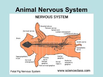 |  |
Nervous System
The nervous system of the pig consists of four basic parts.
The brain - The nervous tissue enclosed by the skull. Part of the central nervous system (CNS). It is covered completely by clear membranes called the meninges.
Spinal cord - The other part of the CNS. It extends from the brain as a narrowed bore tube, through the spinal canal to the tail. Between each of the vertebra, which make up the spine itself, it sends branches out to different parts of he body. The spinal cord is responsible for transmitting the electrical impulses from the brain to these branches.
Peripheral nervous system - Nerves leave the brain and the spinal cord and transmit the electrical impulses throughout the body. This system is the voluntary one that is under the pig's control.
Autonomic nervous system - This is the involuntary nervous system of the pig with separate nerves controlling a wide range of involuntary functions. This system partly controls the heart beat, movement of the muscular walls of the digestive system, the hormonal systems and the excretory systems.
There are a number of important bacterial and viral diseases that cause clinical nervous signs in the pig. Such signs arise by infection of the brain, the brain covering, the spinal cord or any of the peripheral nerves. Some common diseases associated with nervous signs include:Click here to learn more about some of these diseases.
African swine fever (ASF).
Aujeszky's disease (AD) Pseudorabies (PR).
Classical swine fever (CSF) - Hog cholerae (HC).
Congenital tremor - caused by an as yet unidentified virus (possibly a circovirus), swine fever or congenital defects.
Hemagglutinating encephalomyelitis virus (HEV) infection.
Iron toxicity.
Middle ear infection.
Oedema disease (bowel oedema).
Poisons - Arsenic, Mercury, Monensin, Organophosphorus compounds.
Salt toxicity - water deprivation
Porcine stress syndrome (PSS).
Splay leg - a disease of piglets at birth.
Streptococcal meningitis (SM).
Teschen or Talfan diseases.
Tetanus.
Terminology
Cerebrospinal fluid - Fluid that circulates around within the brain and spinal cord. Samples of this fluid can be obtained by needle and syringe for laboratory tests to diagnose nervous disease.
Congenital tremor - A condition in newborn piglets characterised by muscle tremors and shaking. (See chapter 8).
Encephalitis - Inflammation of the brain.
Encephalomyelitis - Inflammation of the brain and spinal cord. Viruses multiplying in the central nervous system primarily cause encephalitis and encephalomyelitis although they may cause meningitis as well. Such viruses include aujeszky's (pseudorabies), rabies, teschen / talfan, haemagglutinating encephalitis (vomiting and wasting disease), classical swine fever, African swine fever, blue eye disease in Mexico, encephalomyocarditis in the US, Caribbean and some other countries, and Japanese B. encephalitis in S.E. Asia.
Meninges - Clear membranes covering the surface of the brain.
Meningitis - Inflammation of the meninges which is extremely painful and often results in dramatic clinical signs. Bacterial infections causing meningitis include Streptococcus suis (mainly type 2), salmonella, Haemophilus parasuis, E. coli and any bacteria gaining access to the meninges from a septicaemia. Viruses may also cause meningitis. Middle ear infection may be mistaken for meningitis.
http://www.thepigsite.com/pighealth/article/7/nervous-system/
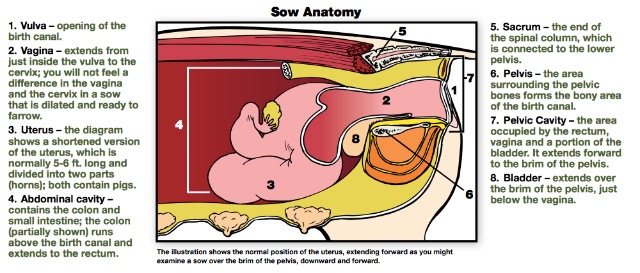
Reproductive Terminology
Abortion - The production of a premature non-viable litter, 111 days or less after mating.
Agalactia - Failure of milk let down or shortage of milk or no milk. The udder may be congested with or without mastitis. In certain conditions, such as mild ergot poisoning, mammary glands fail to develop.
Breeding- The act of mating a sow and boar.
Cervix - The neck of the womb. Inflammation of the cervix is called cervicitis. Cervicitis is not common in the pig, but erosion of the thick folds occurs in old sows and can cause infertility.
Conception rate - As a % this is calculated by: No. of females which conceived x 100 & No. of females mated or inseminated
The calculation is based on a given period of time. Often pig farmers calculate this over the same period of time for both. The top and bottom numbers should be calculated over equivalent periods of time so that the same sows are being counted.
The number conceived can be assessed by the number that did not return to heat or by the numbers judged to be pregnant at the first pregnancy test. Note that the conception rate is generally higher than the farrowing rate because of later embryo / foetus loss. Rolling one month, three month and six month averages of conception rates give an early indication of any developing infertility problems.
Conceptus - Fertilised ovum and embryo.
Corpora haemorrhagica - When the follicle ruptures to release the egg there is a small amount of haemorrhage. This is the name given to the bloody tissues that remain.
Corpus albucans - After pregnancy or after the animal has been in oestrus the corpus luteum disappears and shrinks to a small white body called the corpus albucans.
Corpus luteum - The corpora haemorrhagica becomes consolidated and forms the corpus luteum. This is the body that produces progesterone, the female hormone that maintains pregnancy.
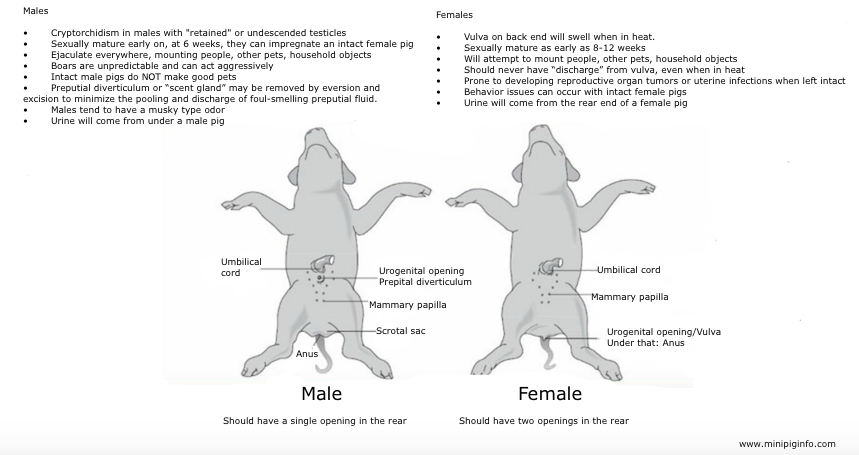
Cryptorchid - A male pig whose testes have not descended through the inguinal canals. Normally, the testes develop in the abdomen and descend through the inguinal canal to the scrotum before birth. Sperm production in the testes require a cooler environment than that of the abdomen.
Embryo - The multicellular organism that develops in the uterus from the fertilised egg up to about 20-30 days when it becomes a foetus.
Endometritis - Inflammation and infection of the lining of the womb (the endometrium).
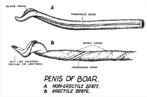
Epididymis - A coiled tube attached to the upper surface of the testicle where the sperm is stored. The sperm leaving it enters the vas deferens. It has a head and a tail. The tail can be cut off (epidectomy) to sterilise the boar.
Erythema - Reddening of the skin that is often seen when one or more mammary glands have mastitis.
Farrowing rate (%) This equals No. females farrowed x 100 or No. females mated
Female - Breeding female including gilts and sows. A gilt becomes a breeding female either from an arbitrary time before mating (e.g. when first brought into the mating area) or, more commonly, when she is first mated. Some pig farmers only include her from the time she farrows but this results in high and less useful indications of herd fertility when farrowing rates and numbers of pigs per female per year are calculated.
Fertilized ovum - The egg as it multiplies and grows to approximately day seven post fertilisation.
Foetus - This describes the developing piglet from approximately 30 days through to maturity.
Inguinal canal - Gap between the muscles of the abdomen in the groin through which the spermatic cord passes from the abdomen to the testicle.
Implantation - The attachment of the embryo to the uterine wall by establishment of the placenta commencing 12 to 14 days post-mating.
Inverted nipples - If the teat sphincter cannot be seen at eye level it is likely that such a teat will remain inverted and will not be functional. This is important to appreciate when selecting or receiving a gilt for breeding. Some inverted nipples will become more normal and be functional when the mammary gland develops but when selecting you cannot take the chance.
Note that each teat has two orifices and teat ducts which drain two quite separate mammary glands, front (anterior) and back (posterior).
Irregular return - A return to oestrus more than 23 days after the previous one.
Lactation length - The period from farrowing to weaning in days.
Litters/per female/per year is calculated by:
Mammary system - The udder of the sow consists of two parallel rows of 5 to 7 teats inter-spaced on each side.
Mastitis - Inflammation of the mammary gland is invariably associated with infection. Bacteria causing it include klebsiella, streptococci, staphylococci and E. coli.
Mastitis metritis agalactia syndrome (MMA) - This syndrome is most commonly associated with mastitis usually coliform mastitis i.e. caused by E. coli or klebsiella but it is also associated with endometritis. The sow is usually sick, running a high temperature and producing little milk. Other terms are sometime used for this syndrome including periparturient hypogalactia syndrome, puerperal toxaemia, and farrowing fever.
Mating - The complete act of copulation involving one or more services.
Mummified pigs - Piglets which died in the uterus and in which the tissues and fluids have been reabsorbed leaving black shrunken skeletal remains.
Non productive days (NPD) - These include all the days when the sows and gilts are either not pregnant or suckling. It therefore always includes:
Oestrus (or heat) - The period during which the sow is receptive to the boar (i.e. will stand to be mated). Usually 1-3 days.
Oestrus cycle - The period from one oestrus to another. 19-22 days interval is normal.
Orchitis - Inflammation of the testicle. A specific example is infection by Brucella suis bacteria. Non infectious orchitis can arise from trauma to one or both testicles. Occasionally there may be a haemorrhage developing into a haematoma (a pocket of blood).
Ovaries - Two small structures which control the oestrus cycle and from which the follicles are produced and the eggs released.
Oxytocin - A hormone produced by the anterior pituitary gland. Its function is to release milk from the glands and at the same time cause the uterus to contract.
Parity - Used to describe the number of times a female has farrowed. e.g.Pregnant gilt = Parity 0; Gilt farrowed for the first time = Parity 1; Sow which has had two litters = Parity 2
(NB. Some people get confused and use the term parity when they mean pregnancy).
Pigs weaned per sow per year - The number of pigs produced in any 12 month period. In a large herd this is usually calculated as a one month, three month and six month rolling average of the whole herd.
Prepucial sac - This is a sac inside the prepuce, the size of a golf ball, that contains a foul smelling fluid with a high bacterial content. Do not squeeze its contents into the vagina at service or you may precipitate an endometritis or cystitis and nephritis. Similarly if you are collecting semen by gloved hand for on-farm artificial insemination you must not contaminate it with prepucial sac contents.
Prolactin - A hormone from the pituitary gland involved in the initiation and maintenance of milk production.
Pyometra - Accumulation of pus in the womb following infection. It is also called pyometritis. This is common when heavy vulva discharges are seen or a retained foetus or placenta are present.
Regular return - A return to oestrus usually 19-22 days after previous one.
Reproductive System- The anatomy of the reproductive tracts of the sow and the boar.
Salpingitis - Inflammation of the oviducts (fallopian tubes) that carry the eggs from the ovary down towards the womb.
Scrotum - This is a sack made of relatively thin pliable skin which has a muscular inner fibro-elastic layer which contracts in a cold environment and relaxes in a hot environment.
Seminal vesicles - These are glands which together with the prostate and bulbo-urethral glands provide fluid and nourishment for the sperm, the fluids being passed out during ejaculation.
Spermatic cord - Fibrous cord, containing the vas deferens and blood vessels, by which the testicles are suspended.
Stillborn pigs - Piglets observed dead behind the sow at birth.
Teat necrosis - Damage to the end of the teat can result in death and sloughing of tissues. This is called necrosis. It is caused by abrasive floor surfaces in the first 18-24 hours of birth and can be an important reason for rejecting gilts for breeding.
Testicle - The gland in which the sperm is produced.
Urethritis - inflammation of the urethra, the tube which carries both sperm and urine down the penis in the boar or urine from the bladder to the vagina in the sow. Urethritis is uncommon in the boar but can occasionally be caused by small calculi or stones formed in the kidneys. The urethra of the sow is much more likely to become contaminated and infected because its opening is so close to the vulva. Urethritis and cystitis are therefore common in the sow.
Uterus (womb) - Consists of two horns upto 1.5m in length that contain the foetuses.
Vagina - The passageway from the exterior to the cervix. Vaginitis (inflammation) occurs following trauma, infection or multiple matings.
Vas deferens - The muscular tube that at ejaculation propels the sperm from the tail of the epididymis on the testes up through the inguinal canal and into the urethra where it joins just below the neck of the bladder . Vasectomising a boar involves cutting the vas deferens midway between the tail of the epididymis and its entry to the abdomen, removing 30-50mm of it.
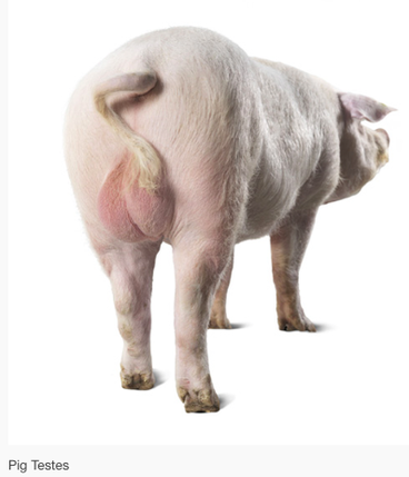
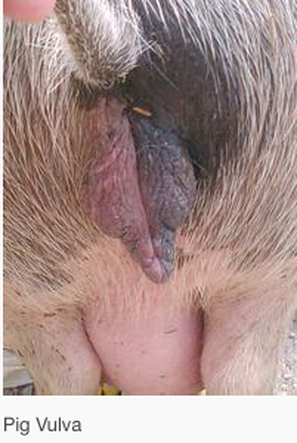
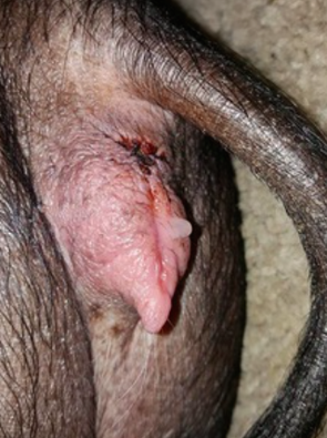
Vulva - The vagina opens to the exterior through the fleshy lips of the vulva. Oedema of the vulva (swelling containing fluid) occurs in late pregnancy and trauma is very common in loose-housed sows. The tissues contain many blood vessels and are prone to hemorrhage. Haemorrhage (haematoma) is also seen in the gilt post farrowing. Such animals can bleed to death.
Source: http://www.thepigsite.com/pighealth/article/8/reproductive-system
Agalactia - Failure of milk let down or shortage of milk or no milk. The udder may be congested with or without mastitis. In certain conditions, such as mild ergot poisoning, mammary glands fail to develop.
Breeding- The act of mating a sow and boar.
Cervix - The neck of the womb. Inflammation of the cervix is called cervicitis. Cervicitis is not common in the pig, but erosion of the thick folds occurs in old sows and can cause infertility.
Conception rate - As a % this is calculated by: No. of females which conceived x 100 & No. of females mated or inseminated
The calculation is based on a given period of time. Often pig farmers calculate this over the same period of time for both. The top and bottom numbers should be calculated over equivalent periods of time so that the same sows are being counted.
The number conceived can be assessed by the number that did not return to heat or by the numbers judged to be pregnant at the first pregnancy test. Note that the conception rate is generally higher than the farrowing rate because of later embryo / foetus loss. Rolling one month, three month and six month averages of conception rates give an early indication of any developing infertility problems.
Conceptus - Fertilised ovum and embryo.
Corpora haemorrhagica - When the follicle ruptures to release the egg there is a small amount of haemorrhage. This is the name given to the bloody tissues that remain.
Corpus albucans - After pregnancy or after the animal has been in oestrus the corpus luteum disappears and shrinks to a small white body called the corpus albucans.
Corpus luteum - The corpora haemorrhagica becomes consolidated and forms the corpus luteum. This is the body that produces progesterone, the female hormone that maintains pregnancy.
Cryptorchid - A male pig whose testes have not descended through the inguinal canals. Normally, the testes develop in the abdomen and descend through the inguinal canal to the scrotum before birth. Sperm production in the testes require a cooler environment than that of the abdomen.
Embryo - The multicellular organism that develops in the uterus from the fertilised egg up to about 20-30 days when it becomes a foetus.
Endometritis - Inflammation and infection of the lining of the womb (the endometrium).
Epididymis - A coiled tube attached to the upper surface of the testicle where the sperm is stored. The sperm leaving it enters the vas deferens. It has a head and a tail. The tail can be cut off (epidectomy) to sterilise the boar.
Erythema - Reddening of the skin that is often seen when one or more mammary glands have mastitis.
Farrowing rate (%) This equals No. females farrowed x 100 or No. females mated
Female - Breeding female including gilts and sows. A gilt becomes a breeding female either from an arbitrary time before mating (e.g. when first brought into the mating area) or, more commonly, when she is first mated. Some pig farmers only include her from the time she farrows but this results in high and less useful indications of herd fertility when farrowing rates and numbers of pigs per female per year are calculated.
Fertilized ovum - The egg as it multiplies and grows to approximately day seven post fertilisation.
Foetus - This describes the developing piglet from approximately 30 days through to maturity.
Inguinal canal - Gap between the muscles of the abdomen in the groin through which the spermatic cord passes from the abdomen to the testicle.
Implantation - The attachment of the embryo to the uterine wall by establishment of the placenta commencing 12 to 14 days post-mating.
Inverted nipples - If the teat sphincter cannot be seen at eye level it is likely that such a teat will remain inverted and will not be functional. This is important to appreciate when selecting or receiving a gilt for breeding. Some inverted nipples will become more normal and be functional when the mammary gland develops but when selecting you cannot take the chance.
Note that each teat has two orifices and teat ducts which drain two quite separate mammary glands, front (anterior) and back (posterior).
Irregular return - A return to oestrus more than 23 days after the previous one.
Lactation length - The period from farrowing to weaning in days.
Litters/per female/per year is calculated by:
- No. of farrowings over 3 months x 4
- Average No. breeding females in the herd
- In large herds this can also be calculated on rolling one month, three month and six month averages which gives a historical indication of rising or falling fertility. Thus for a three month average:
- Average No. females in the herd over last 3 months x 4
- Number of farrowings over last 3 months
Mammary system - The udder of the sow consists of two parallel rows of 5 to 7 teats inter-spaced on each side.
Mastitis - Inflammation of the mammary gland is invariably associated with infection. Bacteria causing it include klebsiella, streptococci, staphylococci and E. coli.
Mastitis metritis agalactia syndrome (MMA) - This syndrome is most commonly associated with mastitis usually coliform mastitis i.e. caused by E. coli or klebsiella but it is also associated with endometritis. The sow is usually sick, running a high temperature and producing little milk. Other terms are sometime used for this syndrome including periparturient hypogalactia syndrome, puerperal toxaemia, and farrowing fever.
Mating - The complete act of copulation involving one or more services.
Mummified pigs - Piglets which died in the uterus and in which the tissues and fluids have been reabsorbed leaving black shrunken skeletal remains.
Non productive days (NPD) - These include all the days when the sows and gilts are either not pregnant or suckling. It therefore always includes:
- Entry of the gilt into the herd to point of mating.
- Time from weaning to mating.
- Time from mating to remating if the female is found not to be pregnant and returns to heat.
- Time after a female has been culled until the time it is slaughtered.
Oestrus (or heat) - The period during which the sow is receptive to the boar (i.e. will stand to be mated). Usually 1-3 days.
Oestrus cycle - The period from one oestrus to another. 19-22 days interval is normal.
Orchitis - Inflammation of the testicle. A specific example is infection by Brucella suis bacteria. Non infectious orchitis can arise from trauma to one or both testicles. Occasionally there may be a haemorrhage developing into a haematoma (a pocket of blood).
Ovaries - Two small structures which control the oestrus cycle and from which the follicles are produced and the eggs released.
Oxytocin - A hormone produced by the anterior pituitary gland. Its function is to release milk from the glands and at the same time cause the uterus to contract.
Parity - Used to describe the number of times a female has farrowed. e.g.Pregnant gilt = Parity 0; Gilt farrowed for the first time = Parity 1; Sow which has had two litters = Parity 2
(NB. Some people get confused and use the term parity when they mean pregnancy).
Pigs weaned per sow per year - The number of pigs produced in any 12 month period. In a large herd this is usually calculated as a one month, three month and six month rolling average of the whole herd.
Prepucial sac - This is a sac inside the prepuce, the size of a golf ball, that contains a foul smelling fluid with a high bacterial content. Do not squeeze its contents into the vagina at service or you may precipitate an endometritis or cystitis and nephritis. Similarly if you are collecting semen by gloved hand for on-farm artificial insemination you must not contaminate it with prepucial sac contents.
Prolactin - A hormone from the pituitary gland involved in the initiation and maintenance of milk production.
Pyometra - Accumulation of pus in the womb following infection. It is also called pyometritis. This is common when heavy vulva discharges are seen or a retained foetus or placenta are present.
Regular return - A return to oestrus usually 19-22 days after previous one.
Reproductive System- The anatomy of the reproductive tracts of the sow and the boar.
Salpingitis - Inflammation of the oviducts (fallopian tubes) that carry the eggs from the ovary down towards the womb.
Scrotum - This is a sack made of relatively thin pliable skin which has a muscular inner fibro-elastic layer which contracts in a cold environment and relaxes in a hot environment.
Seminal vesicles - These are glands which together with the prostate and bulbo-urethral glands provide fluid and nourishment for the sperm, the fluids being passed out during ejaculation.
Spermatic cord - Fibrous cord, containing the vas deferens and blood vessels, by which the testicles are suspended.
Stillborn pigs - Piglets observed dead behind the sow at birth.
Teat necrosis - Damage to the end of the teat can result in death and sloughing of tissues. This is called necrosis. It is caused by abrasive floor surfaces in the first 18-24 hours of birth and can be an important reason for rejecting gilts for breeding.
Testicle - The gland in which the sperm is produced.
Urethritis - inflammation of the urethra, the tube which carries both sperm and urine down the penis in the boar or urine from the bladder to the vagina in the sow. Urethritis is uncommon in the boar but can occasionally be caused by small calculi or stones formed in the kidneys. The urethra of the sow is much more likely to become contaminated and infected because its opening is so close to the vulva. Urethritis and cystitis are therefore common in the sow.
Uterus (womb) - Consists of two horns upto 1.5m in length that contain the foetuses.
Vagina - The passageway from the exterior to the cervix. Vaginitis (inflammation) occurs following trauma, infection or multiple matings.
Vas deferens - The muscular tube that at ejaculation propels the sperm from the tail of the epididymis on the testes up through the inguinal canal and into the urethra where it joins just below the neck of the bladder . Vasectomising a boar involves cutting the vas deferens midway between the tail of the epididymis and its entry to the abdomen, removing 30-50mm of it.
Vulva - The vagina opens to the exterior through the fleshy lips of the vulva. Oedema of the vulva (swelling containing fluid) occurs in late pregnancy and trauma is very common in loose-housed sows. The tissues contain many blood vessels and are prone to hemorrhage. Haemorrhage (haematoma) is also seen in the gilt post farrowing. Such animals can bleed to death.
Source: http://www.thepigsite.com/pighealth/article/8/reproductive-system
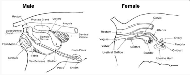
Male Anatomy
  |   |
Female Anatomy
 |  |
Immune System
The various mechanisms that protect the pig from infectious agents can be considered in six groups:
- Complement system - This is a non specific protective mechanism that acts on any foreign cells or viruses that do not possess certain pig proteins on their surface. It consists of a number of chemicals found in the plasma which act together as a cascade to remove or destroy organisms.
- Chemical factors - These include non specific enzymes (such as lysozyme in saliva) and acids which may be found in mucus, saliva and gastric juices. These immobilise or kill pathogens.
- Mechanical factors - These include the skin, mucus, sweat, lining of the nose, mouth, oesophagus, intestine, colon, vagina, flow of urine and the passage of faeces.
- Macrophage cells - These are found throughout the body in tissues and in the blood stream where they are called monocytes. They engulf and digest bacteria. They also have an important role in controlling viral and fungal diseases. The cells are of two types called leucocytes and monocytes.
- Specific acquired immunity - This is of two types; that which is activated by cells and called cell mediated immunity and antibodies present in the blood called humoral immunity. Cell mediated immunity arises when T type lymphocytes come into contact with antigens and they are stimulated to produce antibodies. It takes 7-14 days for these to develop. Humoral immunity is produced from B lymphocytes which have met the antigen previously and their response is immediate. Some lymphocytes also kill other cells that contain antigens or they may act immediately against antigens.
- Immunoglobulins - Specific antibodies of which there are different types namely immunoglobulins, IgG, IgM and IgA. They are found in blood, in milk and particularly in colostrum. All internal surfaces of the body also contain them.
Terminology
Adjuvant - A substance added to an inactivated vaccine to make it more effective.
Antibodies - Complex large proteins (called gamma-globulins) which are produced by specialised cells in response to invading antigens. These stick specifically to the invading antigen neutralising it or triggering off a destructive reaction.
Antigen - Foreign invading substance (i.e. a substance which is not normally part of the pig's body), usually consisting of protein or part of a protein, which stimulates the body to produce antibodies. Antigens exist on the surfaces of bacteria, viruses and parasites.
Antiserum - Serum with high antibody levels against a specific infection. It has usually been produced experimentally in laboratory animals by injecting the infection into them.
Blood sample - Whole blood sample taken hygienically with a syringe into a bottle or by a pin prick through the skin absorbing the droplet of blood with blotting paper.
Commensal bacteria - Bacteria that live permanently in or on the body without causing disease.
Epithelium - Cellular membrane (e.g. mucous membranes) containing epithelial and other cells.
Hyperimmune antiserum - The same as antiserum above but emphasising its high titre.
Lymphocytes - Specialised defence cells in lymph nodes, other lymphatic tissue and the blood which produce antibodies or take part in cellular immunity.
Mucous membranes - Cellular membranes (e.g. those lining the gut) which secrete a sticky substance called mucus on to their surfaces.
Mucous - A clear sticky semi-liquid secreted by cells in mucous membranes.
Pathogenic infection - An infectious organism which has the potential to cause disease. This is in contrast to the many organisms that live normally in or on the body which never cause disease and are called commensals.
Phagocytes - Cells of the body whose special task is to engulf bacteria, viruses, or parasites in an attempt to destroy them. They are also called macrophages.
Phagocytosis - The process whereby the specialised cells of the body engulf bacteria, viruses or parasites in an attempt to destroy them.
Plasma sample - A whole blood sample taken hygienically with a syringe and mixed with an anti-clotting agent so that it remains liquid. The sample is spun fast in a centrifuge and the red and white blood cells sediment to a firm pellet at the bottom leaving a clear liquid - the plasma.
Serology - Tests done in the laboratory to detect the level of specific antibodies in serum samples. ("ology" means study of - so literally serology means "study of serum").
Serum sample - A whole blood sample taken hygienically with a syringe and allowed to clot. The serum is the clear straw-coloured liquid which can be drawn of with a pipette. It contains the antibodies.
Titre - The concentration of a specific antibody in a serum sample. It is expressed as the amount by which the serum has to be diluted before a serological test goes negative.
Virulence - How pathogenic an organism is. Organisms with a high capability of causing disease are called highly virulent.
Source: http://www.thepigsite.com/pighealth/immune-system
Endocrine System
Endocrines or hormones are the substances produced by various glands, which are carried by blood or other body fluids to influence and control the pigs metabolism. There are nine main glands (In picture below) in the pig which are responsible for controlling a variety of vital functions.
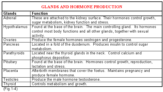
Generally the diseases associated with the failure of the endocrine glands are not important in the pig. However when the regulatory and stimulatory mechanisms between the hypothalamus, the anterior pituitary gland and the ovaries fail, anoestrus (not coming on heat) or reproductive malfunction result, including cystic ovaries. In the male testicular function is affected. The hypothalamus stimulates the anterior part of the pituitary gland to release the follicle stimulating and luteinising hormones (FSH and LH). These in turn act upon the ovaries and the testes to regulate their function.
Terminology
Follicle stimulating hormone (FSH) - Produced by the anterior pituitary gland. It stimulates the formation of follicles in the ovaries,
Growth hormone - Responsible for promoting growth of most tissues throughout the body. It is produced by the pituitary gland in association with the hypothalamus.
Hypothalamus - An area in the brain responsible for providing both nervous and hormonal control over most other hormone producing glands.
Luteinising hormone (LH) - Stimulates ovulation and is produced by the pituitary gland.
Oestrogen - The female hormone responsible for all the female sexual characteristics. It is produced by the ovary.
Oxytocin - Produced by the pituitary gland. This stimulates uterine contractions during farrowing and causes milk let down. It also aids in the movement of sperms and eggs.
Progesterone - The hormone that maintains pregnancy. It is produced by the corpus luteum in the ovary.
Prolactin - This is produced by the pituitary gland and controls milk production.
Prostaglandins - These are produced by the uterus and the placenta and are associated with the initiation of farrowing or abortion.
Testosterone - The male hormone responsible for all the male sexual characteristics. It also controls the development of sperm.
Source: http://www.thepigsite.com/pighealth/endocrine-system
Digestive System
The digestive tract can be considered as a tube that starts at the mouth and finishes at the rectum (Pictured below). In some respect its contents can be considered as outside the body. The back of the mouth opens into the pharynx which is the common area for the passage of both food and air. A valve or flap of tissue called the soft palate automatically moves to protect the opening into the trachea or windpipe when swallowing. The tonsils of the pig are situated on the surface of the soft palate. The oesophagus is the tube that leads from the pharynx to the stomach, down which food is propelled.The main infectious diseases of the mouth are the vesicular ones including foot-and-mouth disease and swine vesicular disease, although occasionally lesions on the skin around the mouth may be seen in aujeszky's disease and porcine reproductive and respiratory syndrome PRRS. Infection of both the gums and bones are common following faulty teeth clipping.
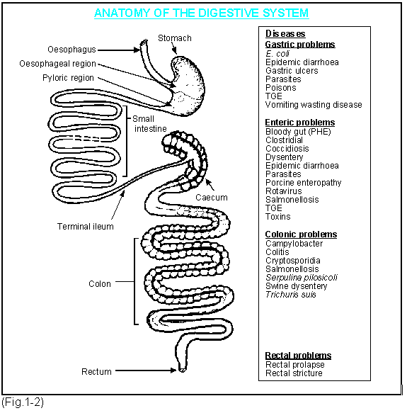
The digestive system of the pig has the ability to convert vegetable and animal materials into highly digestible nutrients. Its anatomy and physiology are similar to that of humans. In the stomach the major disease problems are associated with inflammation of its lining called gastritis which may result in vomiting. Vomiting also occurs in systemic disease where the organism has spread throughout the body (in infections such as erysipelas), and from toxins produced by bacteria or during high fevers.
Gastric ulceration is common in growing pigs occurring in the area where the oesophagus enters the stomach (oesophageal region).
The intestine has two distinctive parts, the small and the large intestine. Inflammation of the former is called enteritis (although sometimes enteritis may mean inflammation of both parts) and the latter colitis. Enteritis is very common and caused by specific viral, bacterial or parasitic infections. The small intestine in cross section contains millions of finger like projections called villi. These increase the absorptive area enormously and thus the efficiency of the digestive process. The large bowel or colon commences with the caecum, the area of the intestinal tract responsible for the digestion of cellulose.
 |  |
Terminology
Ascites - Fluid in the abdomen.
Atrophy - A loss of tissue due to disease or malfunction. Atrophy of the villi in the intestine occurs at weaning time causing malabsorption.
Bloody gut - A descriptive term applied to haemorrhage in the lower part of the small intestine or the complete digestive tract. The latter is seen where there is complete torsion of the intestines. Porcine enteropathy is a common cause. (See chapter 9).
Carbohydrates - These consist of two types, crude fibre and soluble carbohydrates. Crude fibre is a mixture of cellulose. Cellulose digestion takes place in the large intestine.
Cecum - A blind sac, at the beginning of the large intestine.
Colitis - Inflammation of the colon or first part of the large bowel. The caecum is often inflamed at the same time (typhlitis). This is a common condition in young growing pigs from 20-60kg weight, caused by nutritional factors and/or infectious agents.
Colon - The spiral part of the large intestine.
Crypts - The bases of the villi.
Duodenum - This is the first part of the small intestine.
Enteritis - Inflammation of the small intestine. This leads to diarrhoea which is common in sucking pigs, weaners and growers.
Enterocytes - Cells at the base or crypts of the villi in the intestine. They multiply and maintain the length of the villi.
Gall bladder - An organ attached to the liver which produces bile that helps in the digestion and absorption of fats.
Gastric ulcers - Erosions of the mucous lining of the stomach occurring mainly in the oesophageal region. Very common and if severe they result in haemorrhage and death.
Gastritis - Inflammation of the stomach lining. Often causes vomiting.
Gingivitis - Inflammation of the gums.
Glossitis - Inflammation of the tongue.
Hepatitis - Inflammation of the liver.
Ileitis - Inflammation of the ileum.
Ileum - The terminal part of the small intestine.
Jejunum - The middle part of the small intestine.
Liver - This organ is the main factory of the body, building new materials and degrading old ones.
Lignin - See carbohydrates.
Lumen - The open space of the small intestine.
Mucosa - The internal lining of the digestive tract. The cells produce mucus which lubricates the surface and also protects against many pathogenic organisms.
Oesophagus - The muscular tube from the pharynx to the stomach.
Omentum - A reflected net-like membrane from the peritoneum that covers the stomach and intestine.
Pancreas - A gland attached to the duodenum by a tube, which produces digestive enzymes and insulin.
Pars oesophagus - The area of the stomach near the entrance of the oesophagus. A common site for the development of ulcers.
Peritoneum - This is the smooth shiny membrane that covers all the surfaces of the abdomen and its contents.
Peritonitis - Inflammation of the peritoneum.
Pharynx - The common passage for food and air at the back of the throat.
Proteins - These are composed of amino acids which contain carbon, hydrogen, oxygen, sulphur, nitrogen and phosphorus. Combinations of different amino acids produce different proteins.
Pyaemia - Invasion of pus producing organisms throughout the body with small abscess formations.
Soft palate - The flap of tissue that separates the trachea and the oesophagus. It contains the tonsils.
Salivary glands - There are three of these called the parotid, mandibular and sublingual glands. They secrete saliva into the mouth.
Tonsillitis - Inflammation of the tonsils.
Tonsils - Two patches of lymphatic tissue at the back of the throat on the soft palate.
Villi - Finger like projections into the lumen of the small intestine.
Source: http://www.thepigsite.com/pighealth/digestive-system
Circulatory System
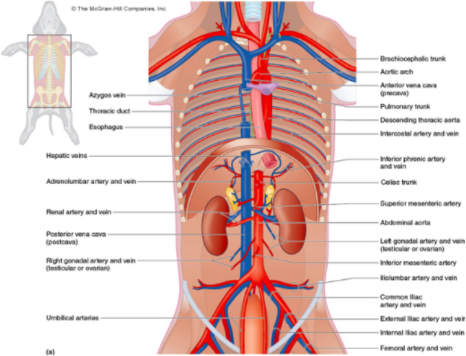
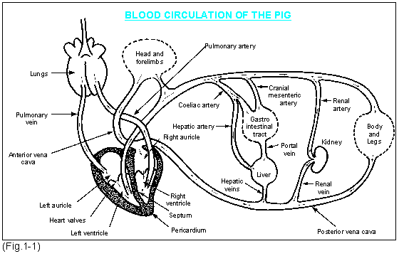
First, study Fig.1-1, then read the following while still referring to the figure. The circulatory system consists of the heart which is a four chamber suction and pressure pump that moves blood through two separate systems, one to and from the lungs and the other around the body. The blood returns to the heart from the body through a series of veins, which terminate in two large veins called the anterior and posterior vena cava. Blood returns from the lungs through the pulmonary veins. The top two chambers or auricles receive the blood from the veins and pass it into the strong muscular bottom chambers called the ventricles. Oxygen depleted blood from the body enters the right auricle, where it is then pumped into the right ventricle leaving by two pulmonary arteries that deliver the still un-oxygenated blood to the lungs. Oxygenated blood from the lungs is then returned through the pulmonary veins to the left auricle, where it is pumped to the left ventricle and finally out through the main artery, the aorta, to be transported around the body. If the lungs are damaged by disease such as pneumonia, they cannot oxygenate the blood efficiently, the tissues become starved of oxygen and cannot function properly.When the pig walks or runs its skin may then become blue and it has difficulty breathing. Chronic pneumonia may also hold back the blood supply causing congestion and heart problems.
Arteries are the muscular tubes that carry the blood away from the heart. These branch off into smaller arteries like the branch of a tree eventually becoming very fine arterioles. The arterioles branch further into microscopic tubes called capillaries which exchange fluid through their walls. This enables the cells of the body to receive both oxygen and nutrients and eliminate carbon dioxide. The capillaries then combine to form first small veins, which in turn lead to larger ones. The blood now contains carbon dioxide and reduced levels of oxygen and returns to the heart via the anterior and posterior vena cava to recommence its circulation around the lung.
There is an important subsidiary circulatory system called the hepatic (i.e. liver) portal system. You will see in Fig.1-1 that two arteries provide oxygen to the stomach and intestines (and also the pancreas and spleen). They keep branching until they form capillaries which then join together to form the portal vein which carries the blood to the liver. There the portal vein breaks up into another capillary-type network, where the blood comes into direct contact with the liver cells. The vessels then join together again to form the hepatic veins which discharge the blood into the posterior vena cava. The blood from the intestines carries nutrients from the food eaten and also sometimes harmful substances (toxins). The liver cells are able to modify some of the nutrients for use elsewhere and also to store some. They also detoxify harmful substances. The liver is supplied with oxygen via a separate artery, the hepatic artery.
The internal linings of the heart are covered by a smooth shiny tissue called the endocardium. The rate of contraction is known as the pulse rate. This can be felt either at the base of the ear or under the tail and varies from 200 beats per minute in the young piglet to 70 in the adult.
The blood consists of two main parts, a fluid called plasma and cells. Nutrients such as proteins, sugars and fats are circulated throughout the body in the plasma and waste products are collected to be detoxified in the liver and excreted via the kidneys. The plasma also carries hormones which are produced in one part of the body and act on another. It also carries antibodies to combat infection. The plasma also supports red blood cells (erythrocytes) which contain the substance haemoglobin whose main function is to transport oxygen around the body and bring back carbon dioxide to be expelled from the lungs. The next largest group in the plasma are the white cells (leucocytes) which are the first line of defence against infectious agents. The third type of cells are blood platelets. These are really small fragments of cells which are associated with the clotting mechanisms of blood. When blood clots the liquid that remains outside the clot is serum and this contains the antibodies. Serum may be used to inject into pigs to provide an immediate source of immunity.
Failure of blood to clot and subsequent loss of red cells into the tissues is not uncommon in pigs and occurs in thrombocytopenic purpura - a clotting defect disease - and warfarin poisoning.
 |  |
Terminology
Albumin - The most abundant protein in the blood.
Anemia - Any reduction in the number of red cells or in the haemoglobin they contain is described as anemia and the extent of this is measured either by determining the number of red cells or the level of haemoglobin in the blood. The causes of anemia include:
Anoxia - Lack of oxygen. Tissues begin to die after a few minutes.
Antibody - The protective proteins produced in response to the antigenic stimulation. They fight infections.
Antigen - This is the foreign protein contained in viruses, bacteria, fungi or toxins. The body responds by producing an antibody.
Antiserum - This is serum containing higher than normal amounts of antibody against a specific antigen. It is used by injection to give an immediate temporary immunity.
Blood count - A laboratory test that determines the numbers of red and white cells and platelets in the blood.
Blood volume - Approximately 8% of body weight expressed as litres .
Blood platelets (thrombocytes) - These are cell fragments involved in blood clotting.
Blood poisoning - A common term used to describe large numbers of pathogenic bacteria in the blood.
Capillaries - Very tiny tubes about the diameter of a red cell. These allow water oxygen and nutrients to diffuse out to the tissues.
Cyanosis - Blueing of the skin and extremities due either to anoxia, toxaemia (toxins in the blood) or septicaemia (pathogenic bacteria in the blood)..
Endocardium - This is the surface tissue lining the inside of the heart. Endocarditis is the end result of the invasion of this tissue by bacteria, in particular erysipelothrix (which causes erysipelas) and streptococci. Both organisms often cause growths on the heart valves called valvular endocarditis. This makes the valves leaky and less effective.
Erythrocytes - These are the red blood cells. In the normal pig there are approximately 7 million per mm3.
Globulins - The proteins that make up the antibodies. They are called gamma globulins.
Granulocytes - These consist of specialised cells called neutrophils, eosinophils and basophils that engulf and destroy bacteria and viruses. They are also called macrophages.
Hematuria - Blood in the urine often seen in cystitis - inflammation of the bladder.
Hemoglobin - This is the chemical substance in the red cells that is involved in the transport of oxygen.
Hemoglobinuria - Free haemoglobulin in the urine resulting from the breakdown of blood cells.
Hemolysis - This is the process by which haemoglobin is released from the red cells when the cell envelope is damaged.
Hydropericardium - Excess fluid around the heart. It is often seen in bacterial infections and shock reactions.
Hypoglycemia - A low level of sugar in the blood. Common in newborn piglets.
Leucocytes - These are the white blood cells of which there are two types, granulocytes and agranulocytes. The granulocytes contain granules in the cell and depending on how they stain they are called neutrophils, eosinophils and basophils. Neutrophils engulf bacteria (phagocytosis), eosinophils increase in chronic disease particularly parasitic disease. Basophils produce a substance called histamine during allergic reactions. Agranulocytes consist of monocytes and lymphocytes.
Lymph - Excessive tissue fluid drained by the lymphatic system. It is similar to plasma.
Lymphatics - A drainage system that removes fluids from tissues and the lymph nodes.
Lymph nodes - These act as filters for lymph and are one of the body's first defences against infection.
Lymphocytes - These are important cells of the immune system producing immunoglobulins. They are of two types, T and B. The total leucocytes in a normal pig are approximately 15,000 per mm3 and numbers increase markedly with bacterial infections. However in some viral diseases their numbers can be significantly reduced.
Macrophages - These take in and usually destroy foreign materials including bacteria and viruses. See granulocytes and monocytes.
Monocytes - These cells engulf bacteria. When they migrate into tissues they become localised tissue macrophages.
Myocardium - Heart muscle.
Myocarditis - Inflammation of the heart muscle. Any scientific term ending with the term "itis" implies inflammation. Inflammation is the body's response to tissue damage and is associated with swelling, poor circulation, reddening, pressure and pain.
Diseases causing myocarditis include streptococcal infections, certain virus infections and deficiencies of Vitamin E or iron. Poisons such as selenium and monensin and the porcine stress syndrome can also cause marked changes to heart muscle.
Oedema - Swelling of tissues due to excess fluid. Common in the udder of the newly farrowed sow.
Oxyhemoglobin - This is haemoglobin combined with oxygen. It is the vehicle by which oxygen is carried around the body.
Pericarditis - The pericardium is the clear sac-like membrane that encloses the heart. Pericarditis occurs as a result of infectious agents which cause respiratory diseases. These include pasteurella, mycoplasma, haemophilus, actinobacillus, streptococci and salmonella bacteria and viruses such as flu and porcine respiratory reproductive virus.
Plasma - Unclotted blood without the blood cells.
Septicemia - Pathogenic bacteria in the blood stream.
Serum - The liquid left after the blood has clotted. It contains large quantities of antibodies which can be used in the laboratory to test for evidence of exposure to diseases or in the field to provide temporary quick protection.
Thrombocyte (blood platelet) - This is responsible for blood clotting.
Thrombosis - The formation of a blood clot in an artery or a vein.
Toxemia - Toxins in the blood stream
Spleen - This organ acts as a reservoir for blood.
Vasiculitis - This describes inflammation of either veins or arteries and it is often a consequence of diseases such as swine fever, erysipelas, Actinobacillus pleuropneumoniae, Haemophilus parasuis and salmonellosis.
Viremia - Viruses in the blood stream.
Source: http://www.thepigsite.com/pighealth/circulatory-system
Albumin - The most abundant protein in the blood.
Anemia - Any reduction in the number of red cells or in the haemoglobin they contain is described as anemia and the extent of this is measured either by determining the number of red cells or the level of haemoglobin in the blood. The causes of anemia include:
- Bowel hemorrhage (proliferative hemorrhagic enteropathy, fungal toxins, acute bowel infection associated with E. coli infection of piglets, salmonella infections or swine dysentery).
- Damage to bone marrow.
- Eperythrozoonosis suis. This is a blood borne bacterium that can destroy red blood cells.
- Gastric ulcers and bleeding - or any other cause of haemorrhage.
- Heavy parasite burdens.
- Iron, copper or vitamin deficiencies.
Anoxia - Lack of oxygen. Tissues begin to die after a few minutes.
Antibody - The protective proteins produced in response to the antigenic stimulation. They fight infections.
Antigen - This is the foreign protein contained in viruses, bacteria, fungi or toxins. The body responds by producing an antibody.
Antiserum - This is serum containing higher than normal amounts of antibody against a specific antigen. It is used by injection to give an immediate temporary immunity.
Blood count - A laboratory test that determines the numbers of red and white cells and platelets in the blood.
Blood volume - Approximately 8% of body weight expressed as litres .
Blood platelets (thrombocytes) - These are cell fragments involved in blood clotting.
Blood poisoning - A common term used to describe large numbers of pathogenic bacteria in the blood.
Capillaries - Very tiny tubes about the diameter of a red cell. These allow water oxygen and nutrients to diffuse out to the tissues.
Cyanosis - Blueing of the skin and extremities due either to anoxia, toxaemia (toxins in the blood) or septicaemia (pathogenic bacteria in the blood)..
Endocardium - This is the surface tissue lining the inside of the heart. Endocarditis is the end result of the invasion of this tissue by bacteria, in particular erysipelothrix (which causes erysipelas) and streptococci. Both organisms often cause growths on the heart valves called valvular endocarditis. This makes the valves leaky and less effective.
Erythrocytes - These are the red blood cells. In the normal pig there are approximately 7 million per mm3.
Globulins - The proteins that make up the antibodies. They are called gamma globulins.
Granulocytes - These consist of specialised cells called neutrophils, eosinophils and basophils that engulf and destroy bacteria and viruses. They are also called macrophages.
Hematuria - Blood in the urine often seen in cystitis - inflammation of the bladder.
Hemoglobin - This is the chemical substance in the red cells that is involved in the transport of oxygen.
Hemoglobinuria - Free haemoglobulin in the urine resulting from the breakdown of blood cells.
Hemolysis - This is the process by which haemoglobin is released from the red cells when the cell envelope is damaged.
Hydropericardium - Excess fluid around the heart. It is often seen in bacterial infections and shock reactions.
Hypoglycemia - A low level of sugar in the blood. Common in newborn piglets.
Leucocytes - These are the white blood cells of which there are two types, granulocytes and agranulocytes. The granulocytes contain granules in the cell and depending on how they stain they are called neutrophils, eosinophils and basophils. Neutrophils engulf bacteria (phagocytosis), eosinophils increase in chronic disease particularly parasitic disease. Basophils produce a substance called histamine during allergic reactions. Agranulocytes consist of monocytes and lymphocytes.
Lymph - Excessive tissue fluid drained by the lymphatic system. It is similar to plasma.
Lymphatics - A drainage system that removes fluids from tissues and the lymph nodes.
Lymph nodes - These act as filters for lymph and are one of the body's first defences against infection.
Lymphocytes - These are important cells of the immune system producing immunoglobulins. They are of two types, T and B. The total leucocytes in a normal pig are approximately 15,000 per mm3 and numbers increase markedly with bacterial infections. However in some viral diseases their numbers can be significantly reduced.
Macrophages - These take in and usually destroy foreign materials including bacteria and viruses. See granulocytes and monocytes.
Monocytes - These cells engulf bacteria. When they migrate into tissues they become localised tissue macrophages.
Myocardium - Heart muscle.
Myocarditis - Inflammation of the heart muscle. Any scientific term ending with the term "itis" implies inflammation. Inflammation is the body's response to tissue damage and is associated with swelling, poor circulation, reddening, pressure and pain.
Diseases causing myocarditis include streptococcal infections, certain virus infections and deficiencies of Vitamin E or iron. Poisons such as selenium and monensin and the porcine stress syndrome can also cause marked changes to heart muscle.
Oedema - Swelling of tissues due to excess fluid. Common in the udder of the newly farrowed sow.
Oxyhemoglobin - This is haemoglobin combined with oxygen. It is the vehicle by which oxygen is carried around the body.
Pericarditis - The pericardium is the clear sac-like membrane that encloses the heart. Pericarditis occurs as a result of infectious agents which cause respiratory diseases. These include pasteurella, mycoplasma, haemophilus, actinobacillus, streptococci and salmonella bacteria and viruses such as flu and porcine respiratory reproductive virus.
Plasma - Unclotted blood without the blood cells.
Septicemia - Pathogenic bacteria in the blood stream.
Serum - The liquid left after the blood has clotted. It contains large quantities of antibodies which can be used in the laboratory to test for evidence of exposure to diseases or in the field to provide temporary quick protection.
Thrombocyte (blood platelet) - This is responsible for blood clotting.
Thrombosis - The formation of a blood clot in an artery or a vein.
Toxemia - Toxins in the blood stream
Spleen - This organ acts as a reservoir for blood.
Vasiculitis - This describes inflammation of either veins or arteries and it is often a consequence of diseases such as swine fever, erysipelas, Actinobacillus pleuropneumoniae, Haemophilus parasuis and salmonellosis.
Viremia - Viruses in the blood stream.
Source: http://www.thepigsite.com/pighealth/circulatory-system
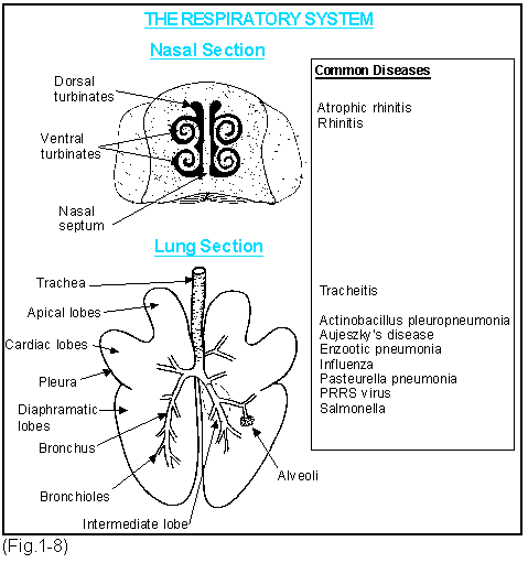
Respiratory System
The respiratory system of the pig commences at the nostrils which lead into two nasal passages. These contain the dorsal and ventral turbinate bones. (see picture below). The ventral turbinates consist of four thin main bones, two on each side separated by a cartilaginous septum. You can imagine these as four hair curlers placed inside the nose. The respiratory tract is lined by a smooth membrane called a mucous membrane because it is bathed in a sticky mucus. It is also covered with minute hair like structures which are able to brush the mucous across the surface by their wavy motion. They move the mucous in the nose, bronchial tree and trachea to the throat where it is swallowed. The air breathed in through the nose is warmed by the turbinate bones which, because of their scroll-like shape, cause turbulence. This throws out the larger of the small particles so that they stick to the mucus and are swept to the throat. The many branches of the bronchi as they decrease in diameter have a similar effect on more minute particles. The mucus elevator then carries them to the throat. Only the vary smallest particles reach the alveoli where the alveolar macrophages engulf and remove them. Internally, the nasal passages open into the pharynx (throat) which is a common passage for food and air. The food is swallowed down the oesophagus and the air is sucked into the larynx at the back of the throat. The larynx (voice box) controls inspiration and expiration. It opens into the trachea which passes down into the chest where it divides into two bronchi. The bronchi branch into smaller bronchi and continue to branch gradually reducing in size to become bronchioles which terminate in very tiny air sacs called alveoli. Oxygen is passed from the alveoli into the blood stream and carbon dioxide is passed out. The lungs are divided into seven lobes as shown in the picture below.
Terminology
Abscess - Area of pneumonia containing pus where the infection has been sealed off from the remainder of the lung tissue by a fibrous capsule.
Actinobacillus pleuropneumoniae - Originally called haemophilus. A bacterium that produces a severe haemorrhagic and necrotising pneumonia with pleurisy.
Alveolar macrophages - These cells which are located in the alveoli engulf bacteria and viruses. They are destroyed by some viruses e.g. the porcine respiratory reproductive syndrome (PRRS) virus.
Atrophic rhinitis - Rhinitis caused by toxigenic (toxin producing) strains of Pasteurella multocida, in which the turbinates loses their tissues (atrophy) irreversibly. This is now called progressive atrophic rhinitis to distinguish it from non-progressive atrophic rhinitis caused by Bordetella bronchiseptica(with the addition of other organisms) and/or environmental contaminants, which is less severe and heals when the infection is stopped by the immune response.
Bronchitis - Inflammation of the bronchi or bronchioles in the lung.
Consolidating pneumonia - The lung tissue has collapsed and become solid. A common example is infection by Mycoplasma hyopneumoniae (enzootic pneumonia) which causes inflammation of the anterior lobes of the lungs.
Enzootic pneumonia - Also called mycoplasmal pneumonia. Caused by Mycoplasma hyopneumoniae, which produces a consolidating pneumonia of the lower parts of the anterior lobes of the lungs.
Glässers disease - Caused by Haemophilus parasuis. It can produce a severe pneumonia and consolidation with fibrinous pleurisy.
Lung worms - Small thread-like worms causing a parasitic pneumonia.
Necrotising pneumonia - Necrosis means death of tissue within the living animal. Necrotising pneumonia occurs where the organism or its toxins kill lung tissue. An abscess may result. A common example is pneumonia caused by Actinobacillus pleuropneumoniae.
Pasteurella - Bacteria found as normal inhabitants of the upper respiratory tract. They often cause secondary infections, for example, following Mycoplasma hyopneumoniae infection. There are two common species P. haemolytica and P. multocidia the latter being the common one in pigs.
Pleurisy - Also called pleuritis. The shiny membranes that covers the surface of the lungs and the inside of the chest wall are called the pleura. Infection or inflammation of these surfaces is called pleurisy. This together with pericarditis is very common in the pig and accounts for considerable loss through condemnation at slaughter. Viruses such as flu, PRRS, swine fever and the bacteria Actinobacillus pleuropneumoniae, Haemophilus parasuis and Pasteurella multocidia can cause pleurisy.
Pneumonia - Inflammation in any part of the lung tissue. There are different types of pneumonia.
Pyaemic pneumonia - Multiple small abscesses scattered through the lungs that have been carried there via the blood stream. A common example is pyaemia from tail biting. The carcase is condemned at slaughter.
Respiratory rate - This varies from 20-40 breaths per minute in piglets and growing pigs and 15-20 per minute in sows.
Rhinitis - Describes any form of inflammation to the delicate mucous lining of the nose. Some agents such as dust and gases may cause it but there is no long-term damage to the nose structure. Sneezing always occurs with rhinitis.
Salmonella choleraesuis - A bacterium specific to the pig causing generalised salmonellosis and pneumonia.
Swine influenza (SI) - A virus infection which produces clearly demarcated dark purple red lesions in the lungs.
Tracheitis - Inflammation of the trachea (windpipe). Influenza may cause a very heavy "barking" cough.
Turbinate bones - Dorsal and ventral. Scrolls of bone inside the nasal passages. They warm and filter air as it passes through the nose.
Source: http://www.thepigsite.com/pighealth/respiratory-system
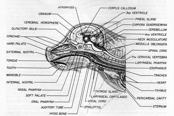
Sensory System
The pig, like the human, experiences sound, sight, smell, taste, heat, cold, pain and balance. The way it responds to these assists us in the recognition of health and disease. For example, pain together with posture will often indicate a specific disease such as fracture of the vertebrae in the spine. Poor balance may be associated with infections of the middle part of the ear which is common in the young growing pig. Likewise; the stockpersons own senses, particularly sight, smell and touch, are important in assessing whether the pig is healthy or ill and performing to its maximum biological efficiency.Sight for example allows the stockperson to observe the lying patterns, any abnormal excretions, signs of disease and unevenness of growth. It also helps to appreciate the quality of the environment.
Smell allows the stockperson to detect toxic gases, blocked drains, putrefying tissue and humidity, important points to consider in respiratory disease. He or she may also learn to detect the smell of scour.
Source: http://www.thepigsite.com/pighealth/sensory-systems
Smell allows the stockperson to detect toxic gases, blocked drains, putrefying tissue and humidity, important points to consider in respiratory disease. He or she may also learn to detect the smell of scour.
Source: http://www.thepigsite.com/pighealth/sensory-systems


My name is Ivan and I have just had the worst years of my life, but now I hope I’m looking at a much
ردحذفbrighter future. This is my story of how my life changed for the worst and how I had to overcome the ignorance of
certain sections of the medical profession. I had to board a plane not once but twice while semi paralysed and fly to
Germany to have the relevant tests done that would prove to all once and for all that I had Lyme/Borreliosis, because
In my own country doctors refused to accept the word of one of their colleagues. The medical profession refused to
consider that I could be suffering from Lyme Disease/Borreliosis even though one of their own had dared to suggest it.
I was watching TV in bed till about 10:30am when I noticed what seemed like the vertical hold slipping and the picture
on the TV going funny for a second. I thought nothing of it at the time and got out of bed and went to make a cup of
tea. Whilst the kettle was on I ran a bath, I was going to clean myself up and go to work to see everyone, because it
was a loss for all the people at work as well as our family. While in the bathroom I felt the floor move just for a
second. I went downstairs thinking that it was odd but nothing more. I made my cup of tea and went up the stairs to
have a good soak in the bath. While drinking my tea and walking up the stairs it happened again, the floor seemed to
move making me take a seat on the stairs, again it soon passed. And I carried on toward the bathroom when my
phone rang in the bedroom - it was work. This was when I noticed that I was slurring on the phone. I hung up and
went to the bathroom to look at myself in the mirror. I looked ok and was feeling ok.
Whatever it was I still didn’t feel it was anything too serious. I got into the bath and then it all started I couldn’t stand.
I felt like gravity was getting it’s own back for all the years I had taken it for granted. I was now fighting to keep my
head above the water while I tried to take the plug out with my left foot as the right one had lost all coordination.
I managed to slide out of the bath, how long it took is all a blur now that I look back. Now I was convinced I was
dying. My children were at school and my wife had gone to see friends, I was alone and scared, my face was numb
down the one side, what was happening to me? Then after what seemed a lifetime but I’m sure was only a few
minutes it started to fade, I managed to drag myself to the bedroom naked and soaked.
Then I got introduce to Dr Itua Herbal Medicine by a friend of mine at work when he sees how I was battling with the disease, He gave me Dr Itua contact so I contacted him on whatsapp then I explain to him on how I was diagnosis of lyme disease so he assure me that he will cure me with his herbal medicine which I will drink for three weeks to cure so I make a purchase with the herbal medicine through moneygram money transfer so faithful of him he deliver me the herbal medicine so he instructed me on how to drink it which I did and hopefully like he promised me the herbal medicine cured me and today I'm now a free person and I thank my dear friend who introduce Dr Itua to my life.
You can contact Dr Itua On Email or whatsapp .... . drituaherbalcenter@gmail.com / Whatsapp ... +2348149277967 if you are suffering from the below disease written because he promised me that he can cure the following diseases......Herpes,Prostate Breast Cancer,Brain Cancer,CEREBRAL VASCULAR ACCIDENT,Endometrial Cancer, cerebrovascular diseases,Hepatitis,Glaucoma,Cataracts,Macular degeneration,Cardiovascular disease,Lung disease.Enlarged prostate,Osteoporosis.Alzheimer's disease,
Dementia.Tach Disease,HIV,Arthritis,Amyotrophic Fibromyalgia,Cervical Cancer,Colon Cancer,Blood Cancer,
SYPHILIS,Diabetes,Liver / Inflammatory kidney,Epilepsy,Shingles,H.P.V TYPE 1 TYPE 2 TYPE 3 AND TYPE 4. TYPE 5.
Lung Cancer, Leukemia Lymphoma Cancer,Lung Mesothelioma Asbestos,Ovarian Cervical Uterine Cancer,Skin Cancer,Brain Tumor.